

It is the inability of the veins (veins) carrying the dirty blood coming out of the testicles in men to not work properly due to the discomfort and therefore not being able to carry the dirty blood inside to the heart. Due to the dirty blood accumulating in these veins, the veins swell and wrinkle. We call varicocele similar to the varicose veins that occur in the testicles in the legs (especially in women).
It is one of the most common and correctable causes of male infertility (male infertility). It is seen in 20% of adult men in the society. It is found in 35% of men who apply with the complaint of not being able to have children.
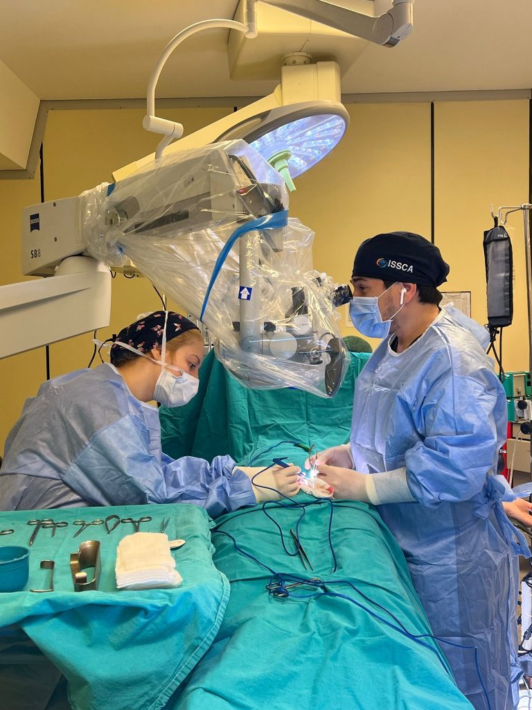
Varicocele is a progressive disease and regression in testicular development; It can cause infertility (inability to have children) by disrupting sperm production. Although it is mostly seen on the left side, it should be investigated thoroughly whether it is bilateral or not.
Varicocele can cause the following adverse conditions that can be revealed by spermiogram (sperm analysis) in the semen:
- Decrease in sperm count
- Impaired sperm motility
- Sperm shape (morphology) deterioration
These adverse effects are caused by the following mechanisms:
- Increase in temperature as a result of the accumulation of blood that cannot be expelled in the testicles
- Harmful products that need to be removed from the testis cannot be removed from the damaged vessels and returned to the testis (reflux).
- Accumulation of dirty blood in the testis
How is varicocele diagnosed (diagnosed)?
The most important method in diagnosis is an examination by an experienced urologist. The examination should be done standing up. Physical examination is the most valuable method. according to inspection
Varicocele is divided into 3 degrees:
- Stage I (stage I): It is the mildest degree and can be palpated during outpatient examination only with maneuvers such as coughing/straining.
- Stage II (grade II): In the outpatient examination, there is no need to increase the intra-abdominal pressure, which can be understood from the manual examination.
- Stage III (grade III): This is the most severe type and there are varicose veins that appear on the feet.
It occurs with the feeling of varicose veins by hand in cases that increase intra-abdominal pressure. The second step in the diagnosis is scrotal color Doppler ultrasonography in the presence of conditions that make physical examination difficult. With this examination, the diameters of the sick veins, whether the dirty blood returns to these veins, and whether there is a loss of volume in the testicles are revealed.
Sperm Analysis (Spermiogram)
Varicocele; It can cause sperm count, movement and deformity (morphology). Ideally, 2 separate sperm analyzes (spermiograms) should be performed in the evaluation of the patient; There should be a minimum of 7 days and a maximum of 3 weeks between two sperm analysis.
The sperm count is 5-10 million. If the patient’s blood is between FSH and Testosterone, it should be checked. If the number is less than 5 million: Genetic tests (Karyotype and Y chromosome) should be performed. If a defect is detected as a result of genetic testing, the varicocele was probably found by chance and varicocele surgery will not benefit these patients.
Subclinical Varicocele
It is a varicocele that the surgeon could not detect in his examination, but revealed by Scrotal Color Doppler Ultrasound. Surgical treatment results of such cases are much lower than high-grade varicocele treatment results.
Pain and Varicocele
Although varicocele is a disease that does not cause any symptoms, but is detected when married couples apply to a urologist due to inability to have children, approximately 6% of patients experience testicular pain and/or discomfort. Pain alone does not require surgery; However, if the pain is not relieved by other methods, surgical treatment may be considered.
Varicocele Surgical Treatment (Surgery)
The current surgical treatment of varicocele is to reach the testicular veins with an incision of approximately 2-3 cm from the inguinal region and to cancel the diseased veins by ligating them. Apart from this classical method, Laparoscopic, Robotic and Radiological Embolization methods have also been applied before, but today they are not used because the desired results cannot be obtained as much as classical surgery.
Since it is not possible to reach all the veins of the testis in laparoscopy, these veins cannot be ligated, which means that the surgical procedure is incomplete. In addition, there are risks such as the possibility of injury to the intra-abdominal organs in laparoscopic and robotic methods.
Radiologically, the method of occlusion (embolization) of the varicose veins was also used for a period, but in most cases, the classical method was used because testicular veins could not be entered. The aim of the surgical treatment of varicocele is to connect the diseased vessels, to protect the artery, sperm duct (vas deferens) and lymph vessels that bring clean blood to the testicles. In the best method applied in the treatment of varicocele; Complication rates such as varicocele recurrence, vascular injury and postoperative hydrocele formation (fluid collection in the testis) should be the lowest, and postoperative sperm parameters should be improved and pregnancy rates should be higher than other methods.
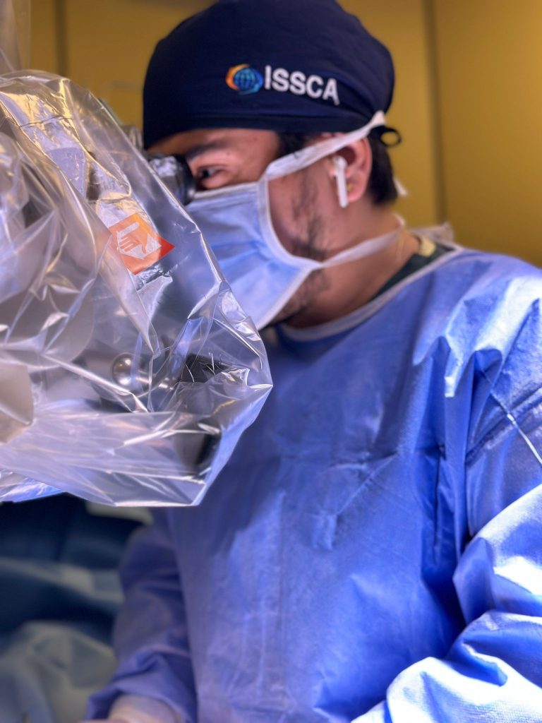
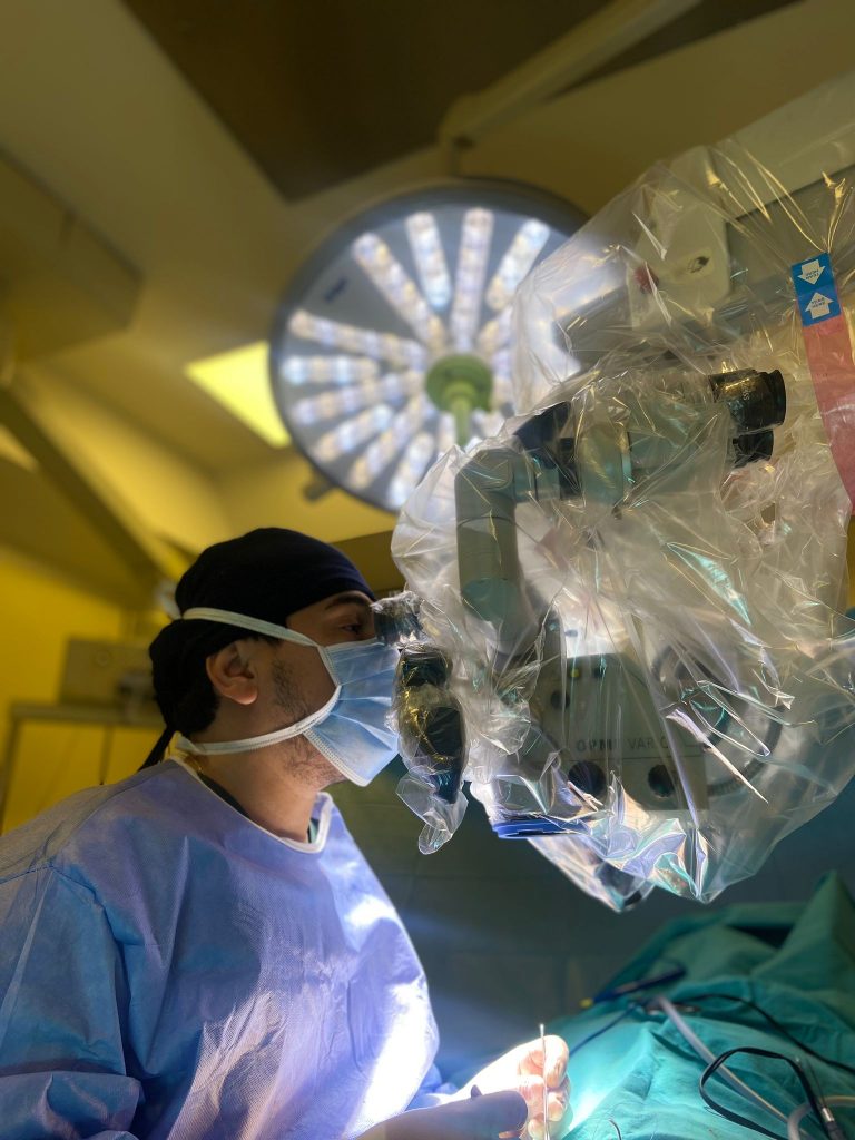
For this reason, this operation should be performed with the “MICRO SURGERY” technique, that is, using the operating microscope. While the probability of recurrence (recurrence) of the disease with the microsurgery method is 1%, this rate is 15% if the operating microscope is not used. In addition, as a result of the operation performed with the microsurgery method, the pregnancy rate in the first year was 43%.


